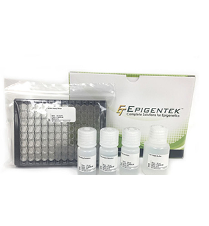
Mast Cell Tryptase (MCT) BioAssay ELISA Kit (Rat) s a 1.5 hour solid-phase Competitive ELISA designed for the quantitative determination of Mast Cell Tryptase (MCT) in rouse serum, plasma, cell culture supernatants, body fluid and tissue homogenates.Detection Range:0.5-10ng/mlSensitivity:0.1ng/mlPrecision:Intra-Assay CV: <10%Inter-Assay CV: <10%Specificity: This assay has high sensitivity and excellent specificity for detection of MCT (Rat). No significant crossreactivity or interference with analogs was observed. Note: Crossreactivity detection between the MCT (Rat) and all analogs has not been determined and crossreaction may exist in some cases.Kit Components:196455A: Microtiter Strips, 1x48 or 1x96 wells196455B: Enzyme Conjugate, 1x3ml or 1x6ml196455C: Standard, 0ng/ml, 1x1 vial196455D: Standard, 0.5ng/ml, 1x1 vial196455E: Standard, 1ng/ml, 1x1 vial196455F: Standard, 2.5ng/ml, 1x1 vial196455G: Standard, 5ng/ml, 1x1 vial196455H: Standard, 10g/ml, 1x1 vial196455J: Substrate A, 1x6ml196455K: Substrate B, 1x6ml196455L: Stop Solution, 1x6ml196455M: Wash Solution (100X), 1x10ml196455N: Balance Solution, 1x3mlStorage and Stability:Store components at 4°C. Stable for 6 months after receipt. For maximum recovery of product, centrifuge the original vial prior to removing the cap.Assay Principle:Mast Cell Tryptase (MCT) BioAssay ELISA Kit (Rat) utilizes the quantitative Competitive Enzyme Immunoassay technique, utilizing a monoclonal antibody and an conjugate to Mast Cell Tryptase (MCT) (Rat). The assay sample and buffer are incubated together with the HRP conjugate in a pre-coated plate for one hour. After the incubation period, the wells are decanted and washed five times. The wells are then incubated with a substrate for HRP enzyme. The product of the enzyme-substrate reaction forms a blue colored complex. Finally, a stop solution is added to stop the reaction, which will then turn the solution yellow. The intensity of color is measured spectrophotometrically at 450nm in a microplate reader. The intensity of the color is inversely proportional to the MCT concentration since MCT from samples and MCT-HRP conjugate compete for the anti-MCT antibody binding site. Since the number of sites is limited, as more sites are occupied by MCT from the sample, fewer sites are left to bind the MCT-HRP conjugate. A standard curve is plotted relating the intensity of the color (O.D.) to the concentration of standards. The MCT concentration in each sample is interpolated from this standard curve.Contact:United States Biological4 Technology WaySalem, MA 01970www.usbio.net