PDL1 belongs to the immunoglobulin superfamily and BTN/MOG family. PDL1 is involved in the costimulatory signal, essential for T-cell proliferation and production of IL-10 and IFNG, in an IL-2 dependent and a PDCD1 independent manner. It is up-regulated in T-and B-cells, dendritic cells, keratinocytes and monocytes after LPS and IFNG activation. PDL1 is up-regulated in B-cells activated by surface Ig cross-linking. Interaction with PDCD1 inhibits T-cell proliferation and cytokine production. During infection or inflammation of normal tissue, this interaction is important for preventing autoimmunity by maintaining homeostasis of the immune response. In tumor microenvironments, this interaction provides an immune escape for tumor cells through cytotoxic T-cell inactivation. Expression of this gene in tumor cells is considered to be prognostic in many types of human malignancies, including colon cancer and renal cell carcinoma. Anti-PDL-1 Antibody is useful for researches interested in cancer research, NF-kB pathway research, and the Immune system.
Anti-PDL1 antibody was prepared from whole goat serum produced by repeated immunizations with a synthetic peptide corresponding to an internal portion of human PDL1 conjugated to Keyhole Limpet Hemocyanin (KLH).
来源宿主
Goat
反应性
H. sapiens (Human); Mus musculus (Mouse)
保存建议
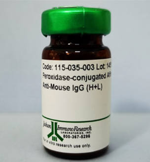
Store vial at -20° C or below prior to opening. This vial contains a relatively low volume of reagent (25 µL). To minimize loss of volume dilute 1:10 by adding 225 µL of the buffer stated above directly to the vial. Recap, mix thoroughly and briefly centrifuge to collect the volume at the bottom of the vial. Use this intermediate dilution when calculating final dilutions as recommended below. Store the vial at -20°C or below after dilution. Avoid cycles of freezing and thawing.